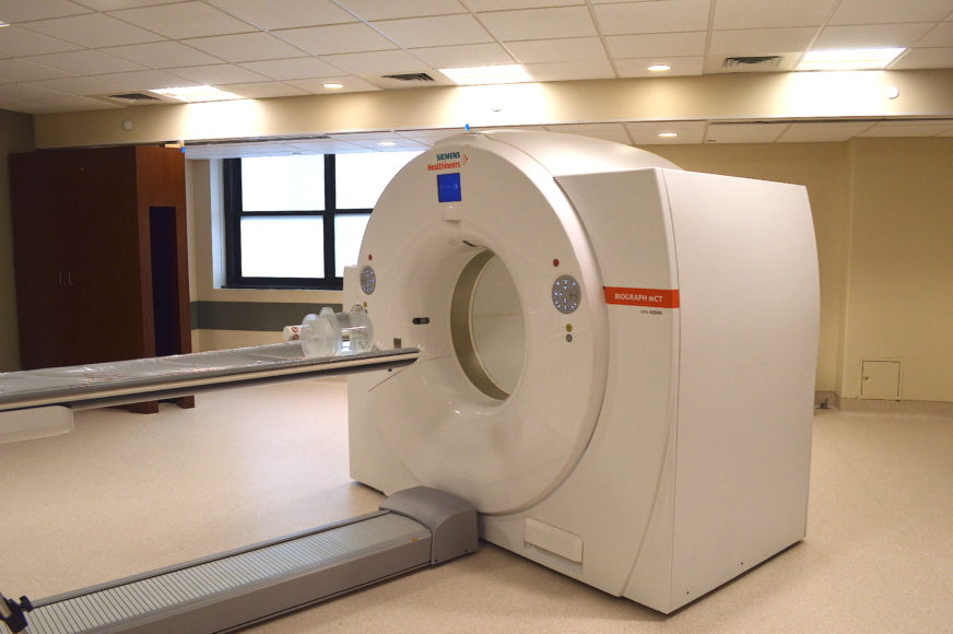
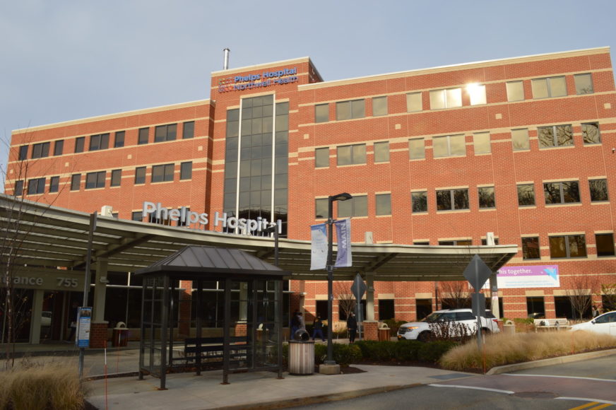
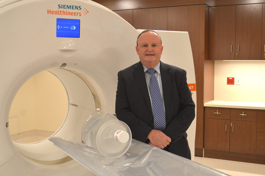
At Phelps Hospital in Sleepy Hollow, turning the page on the calendar to the new year meant looking forward to the launch of the new $8.4 million high-tech imaging suite this month. The centerpiece of the facility is a new PET/CT scanner built by Semens Healthineers. The machine simultaneously does Positron Emission Tomography (PET) and Computerized Tomography Imaging (CT) imaging. Obtaining both PET and CT images previously required two separate sessions using two separate pieces of equipment.
Phelps is a Northwell Health hospital and the new 3,800-square-foot imaging suite will be used by patients at the Northwell Health Cancer Institute at Phelps in addition to other patients being diagnosed and treated for issues such as cardiac problems, seizures and dementia. The new imaging suite is located on the first floor of the hospital, not far from the lobby at the hospital’s main entrance.
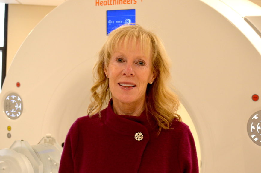
“It’s very exciting because we’re able to fully enhance our radiological capabilities here, our imaging studies, but even more important we’re going to be able to add to our cancer center and be able to provide care for our cancer patients that we otherwise weren’t able to,” Eileen Egan, executive director of Phelps, tells WAG.
When ordinary X-rays or an MRI scan are not able to help physicians pinpoint problems affecting patients, PET/CT imaging may be useful. Egan says that one of the things the staff at Phelps is proudest of is that the new equipment makes it possible for patients needing PET/CT imaging no longer have to go into Manhattan or travel elsewhere to find an imaging source.
“It’s an advanced level of imaging that really may help identify any problems that may otherwise not have been picked up by other imaging modalities,” she says. “The equipment is very expensive, but we anticipate with the volume of patients that we’re going to be able to see and treat we know it’s going to pay for itself very quickly.”
She says that a constant goal when designing the new imaging center was patient comfort.
“If they require this type of imaging, we want them to feel comfortable in the environment,” she adds. “This space is very spacious. It’s open, there’s light, there are windows. We want our patients not to feel claustrophobic when they’re having their testing done.”
Egan says that the new imaging center coincides with other efforts at Phelps to expand the services it offers to the community.
“We are proud to be developing a Center for Advanced Procedures, which is going to be focused on neurosciences and neuroendovascular cases, so this is all part of that,” she says, referring to diseases of the nervous system and its blood vessels. “But this particularly is part of our plans to develop a comprehensive cancer center in Westchester on the Phelps campus, and this will greatly enhance our oncologists’ ability to diagnose and treat cancer.”
Michael Glennon, assistant vice president, operations, ancillary services at Phelps, tells WAG: “I’ve been in radiology most of my life. I actually started out hand-developing X-ray films in tanks. Now, it’s all electronic, all digital. Everything can be sent and seen anywhere.”
Glennon explains that with digital files rather than pieces of physical film you can have an expert on Long Island or elsewhere at Northwell instantaneously see the images and collaborate in developing a diagnosis or treatment plan.
In the imaging center, adjusted lighting and added music can help the patient relax. Glennon points out that the scanner uses a wide bore, or opening into which the patient is moved, to help reduce patient anxiety and overcome the claustrophobia that patients had with earlier scanners, which put them into a tight tunnel.
“Our technologists already know the CT scan technology and we’ve hired another nuclear medicine technologist who knows the PET side of it, so combined there will be two experts in the room always working the equipment,” Glennon says. “You’re getting a radiopharmaceutical injection (which emits radiation picked up by the imaging machine) in your arm and then you lie on the table. It’s very easy. It used to be over an hour. It’s down to about 20 minutes now for most procedures.”
Glennon explains that where standard X-rays show shadows, the PET/CT scans show not only distinct images but also the chemical makeup because the radiopharmaceutical agents actually attach to different cells within the body and thus show up in different colors and intensities.
“We can see the intensity; we can see the width, the depth, 3D. It’s all calculated,” Glennon says. “We’ve come through so many levels of technology over the years. This is quite amazing. I never thought we’d be here. When you see the 3D imaging, it’s quite amazing.”
For more, visit phelps.northwell.edu.